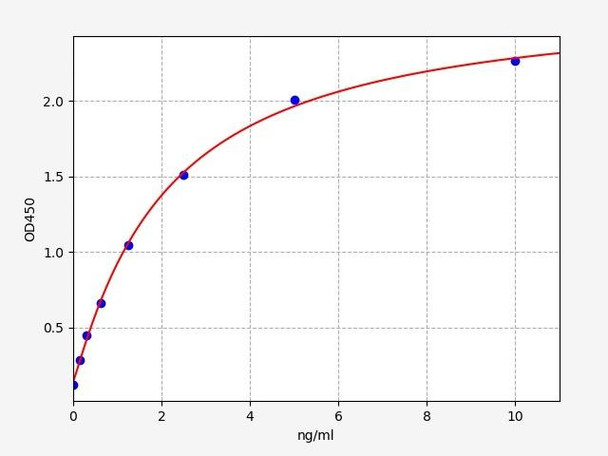
Human Ephrin A5 ELISA Kit
- SKU:
- HUFI02410
- Product Type:
- ELISA Kit
- Size:
- 96 Assays
- Uniprot:
- P52803
- Sensitivity:
- 0.094ng/ml
- Range:
- 0.156-10ng/ml
- ELISA Type:
- Sandwich ELISA, Double Antibody
- Synonyms:
- EFNA5, AL-1, EFL-5, LERK-7, RAGS, AF1, AL-1, EPH-related receptor tyrosine kinase ligand 7, EPLG7, GLC1M, LERK-7, LERK7EFL5, RAGS
- Reactivity:
- Human
Description
| 商品名: | Human Ephrin A5 ELISA Kit |
| 製品コード: | HUFI02410 |
| サイズ: | 96T |
| エイリアス: | EFNA5, AL-1, EFL-5, LERK-7, RAGS, AF1, AL-1, EPH-related receptor tyrosine kinase ligand 7, EPLG7, GLC1M, LERK-7, LERK7EFL5, RAGS |
| 検出方法: | Sandwich ELISA, Double Antibody |
| 申し込み: | This immunoassay kit allows for the in vitro quantitative determination of Human EFNA5 concentrations in serum plasma and other biological fluids. |
| 感度: | 0.094ng/ml |
| 範囲: | 0.156-10ng/ml |
| 保管所: | 4°C for 6 months |
| ノート: | For Research Use Only |
| 回復: | Matrices listed below were spiked with certain level of Human EFNA5 and the recovery rates were calculated by comparing the measured value to the expected amount of Human EFNA5 in samples. | ||||||||||||||||
| |||||||||||||||||
| 直線性: | The linearity of the kit was assayed by testing samples spiked with appropriate concentration of Human EFNA5 and their serial dilutions. The results were demonstrated by the percentage of calculated concentration to the expected. | ||||||||||||||||
| |||||||||||||||||
| CV(%): | Intra-Assay: CV<8% Inter-Assay: CV<10% |
| 成分 | 量 | 保管所 |
| ELISA Microplate (Dismountable) | 8×12 strips | 4°C for 6 months |
| Lyophilized Standard | 2 | 4°C/-20°C |
| Sample/Standard Dilution Buffer | 20ml | 4°C |
| Biotin-labeled Antibody(Concentrated) | 120ul | 4°C (Protect from light) |
| Antibody Dilution Buffer | 10ml | 4°C |
| HRP-Streptavidin Conjugate(SABC) | 120ul | 4°C (Protect from light) |
| SABC Dilution Buffer | 10ml | 4°C |
| TMB Substrate | 10ml | 4°C (Protect from light) |
| Stop Solution | 10ml | 4°C |
| Wash Buffer(25X) | 30ml | 4°C |
| Plate Sealer | 5 | - |
必要なその他の材料と設備:
- Microplate reader with 450 nm wavelength filter
- Multichannel Pipette, Pipette, microcentrifuge tubes and disposable pipette tips
- Incubator
- Deionized or distilled water
- Absorbent paper
- Buffer resevoir
| Uniprot | P52803 |
| UniProt Protein Function: | EFNA5: Cell surface GPI-bound ligand for Eph receptors, a family of receptor tyrosine kinases which are crucial for migration, repulsion and adhesion during neuronal, vascular and epithelial development. Binds promiscuously Eph receptors residing on adjacent cells, leading to contact-dependent bidirectional signaling into neighboring cells. The signaling pathway downstream of the receptor is referred to as forward signaling while the signaling pathway downstream of the ephrin ligand is referred to as reverse signaling. Induces compartmentalized signaling within a caveolae-like membrane microdomain when bound to the extracellular domain of its cognate receptor. This signaling event requires the activity of the Fyn tyrosine kinase. Activates the EPHA3 receptor to regulate cell-cell adhesion and cytoskeletal organization. With the receptor EPHA2 may regulate lens fiber cells shape and interactions and be important for lens transparency maintenance. May function actively to stimulate axon fasciculation. The interaction of EFNA5 with EPHA5 also mediates communication between pancreatic islet cells to regulate glucose-stimulated insulin secretion. Cognate/functional ligand for EPHA7, their interaction regulates brain development modulating cell-cell adhesion and repulsion. Belongs to the ephrin family. |
| UniProt Protein Details: | Protein type:Cell development/differentiation; Ligand, receptor tyrosine kinase; Motility/polarity/chemotaxis; Membrane protein, GPI anchor Chromosomal Location of Human Ortholog: 5q21 Cellular Component: anchored to external side of plasma membrane; extracellular region; plasma membrane; caveola Molecular Function:ephrin receptor binding; chemorepellent activity Biological Process: nervous system development; axon guidance; positive regulation of peptidyl-tyrosine phosphorylation; regulation of cell-cell adhesion; apoptosis; regulation of actin cytoskeleton organization and biogenesis; regulation of focal adhesion formation; ephrin receptor signaling pathway; negative chemotaxis; retinal ganglion cell axon guidance |
| NCBI Summary: | Ephrin-A5, a member of the ephrin gene family, prevents axon bundling in cocultures of cortical neurons with astrocytes, a model of late stage nervous system development and differentiation. The EPH and EPH-related receptors comprise the largest subfamily of receptor protein-tyrosine kinases and have been implicated in mediating developmental events, particularly in the nervous system. EPH receptors typically have a single kinase domain and an extracellular region containing a Cys-rich domain and 2 fibronectin type III repeats. The ephrin ligands and receptors have been named by the Eph Nomenclature Committee (1997). Based on their structures and sequence relationships, ephrins are divided into the ephrin-A (EFNA) class, which are anchored to the membrane by a glycosylphosphatidylinositol linkage, and the ephrin-B (EFNB) class, which are transmembrane proteins. The Eph family of receptors are similarly divided into 2 groups based on the similarity of their extracellular domain sequences and their affinities for binding ephrin-A and ephrin-B ligands. [provided by RefSeq, Jul 2008] |
| UniProt Code: | P52803 |
| NCBI GenInfo Identifier: | 1706678 |
| NCBI Gene ID: | 1946 |
| NCBI Accession: | P52803.1 |
| UniProt Related Accession: | P52803 |
| Molecular Weight: | 26,297 Da |
| NCBI Full Name: | Ephrin-A5 |
| NCBI Synonym Full Names: | ephrin-A5 |
| NCBI Official Symbol: | EFNA5 |
| NCBI Official Synonym Symbols: | AF1; EFL5; RAGS; EPLG7; GLC1M; LERK7 |
| NCBI Protein Information: | ephrin-A5; AL-1; LERK-7; eph-related receptor tyrosine kinase ligand 7 |
| UniProt Protein Name: | Ephrin-A5 |
| UniProt Synonym Protein Names: | AL-1; EPH-related receptor tyrosine kinase ligand 7; LERK-7 |
| Protein Family: | Ephrin |
| UniProt Gene Name: | EFNA5 |
| UniProt Entry Name: | EFNA5_HUMAN |
*ノート: プロトコルは、各バッチ/ロットに固有です。正しい手順については、キットに含まれているプロトコルに従ってください。
ウェルに加える前に、SABCワーキング溶液とTMB基質を37°Cで少なくとも30分間平衡化します。 サンプルと試薬を希釈するときは、完全に均一に混合する必要があります。各テストの標準曲線をプロットすることをお勧めします。
| ステップ | プロトコル |
| 1. | Set standard, test sample and control (zero) wells on the pre-coated plate respectively, and then, record their positions. It is recommended to measure each standard and sample in duplicate. Wash plate 2 times before adding standard, sample and control (zero) wells! |
| 2. | Aliquot 0.1ml standard solutions into the standard wells. |
| 3. | Add 0.1 ml of Sample / Standard dilution buffer into the control (zero) well. |
| 4. | Add 0.1 ml of properly diluted sample ( Human serum, plasma, tissue homogenates and other biological fluids.) into test sample wells. |
| 5. | Seal the plate with a cover and incubate at 37 °C for 90 min. |
| 6. | Remove the cover and discard the plate content, clap the plate on the absorbent filter papers or other absorbent material. Do NOT let the wells completely dry at any time. Wash plate X2. |
| 7. | Add 0.1 ml of Biotin- detection antibody working solution into the above wells (standard, test sample & zero wells). Add the solution at the bottom of each well without touching the side wall. |
| 8. | Seal the plate with a cover and incubate at 37°C for 60 min. |
| 9. | Remove the cover, and wash plate 3 times with Wash buffer. Let wash buffer rest in wells for 1 min between each wash. |
| 10. | Add 0.1 ml of SABC working solution into each well, cover the plate and incubate at 37°C for 30 min. |
| 11. | Remove the cover and wash plate 5 times with Wash buffer, and each time let the wash buffer stay in the wells for 1-2 min. |
| 12. | Add 90 µl of TMB substrate into each well, cover the plate and incubate at 37°C in dark within 10-20 min. (Note: This incubation time is for reference use only, the optimal time should be determined by end user.) And the shades of blue can be seen in the first 3-4 wells (with most concentrated standard solutions), the other wells show no obvious color. |
| 13. | Add 50 µl of Stop solution into each well and mix thoroughly. The color changes into yellow immediately. |
| 14. | Read the O.D. absorbance at 450 nm in a microplate reader immediately after adding the stop solution. |
ELISAアッセイを実施する場合、可能な限り最良の結果を達成するためにサンプルを準備することが重要です。以下に、さまざまなサンプルタイプのサンプルを準備するための手順のリストを示します。
| サンプルタイプ | プロトコル |
| 血清 | If using serum separator tubes, allow samples to clot for 30 minutes at room temperature. Centrifuge for 10 minutes at 1,000x g. Collect the serum fraction and assay promptly or aliquot and store the samples at -80°C. Avoid multiple freeze-thaw cycles. If serum separator tubes are not being used, allow samples to clot overnight at 2-8°C. Centrifuge for 10 minutes at 1,000x g. Remove serum and assay promptly or aliquot and store the samples at -80°C. Avoid multiple freeze-thaw cycles. |
| プラズマ | Collect plasma using EDTA or heparin as an anticoagulant. Centrifuge samples at 4°C for 15 mins at 1000 × g within 30 mins of collection. Collect the plasma fraction and assay promptly or aliquot and store the samples at -80°C. Avoid multiple freeze-thaw cycles. Note: Over haemolysed samples are not suitable for use with this kit. |
| 尿および脳脊髄液 | Collect the urine (mid-stream) in a sterile container, centrifuge for 20 mins at 2000-3000 rpm. Remove supernatant and assay immediately. If any precipitation is detected, repeat the centrifugation step. A similar protocol can be used for cerebrospinal fluid. |
| 細胞培養上清 | Collect the cell culture media by pipette, followed by centrifugation at 4°C for 20 mins at 1500 rpm. Collect the clear supernatant and assay immediately. |
| 細胞溶解物 | Solubilize cells in lysis buffer and allow to sit on ice for 30 minutes. Centrifuge tubes at 14,000 x g for 5 minutes to remove insoluble material. Aliquot the supernatant into a new tube and discard the remaining whole cell extract. Quantify total protein concentration using a total protein assay. Assay immediately or aliquot and store at ≤ -20 °C. |
| 組織ホモジネート | The preparation of tissue homogenates will vary depending upon tissue type. Rinse tissue with 1X PBS to remove excess blood & homogenize in 20ml of 1X PBS (including protease inhibitors) and store overnight at ≤ -20°C. Two freeze-thaw cycles are required to break the cell membranes. To further disrupt the cell membranes you can sonicate the samples. Centrifuge homogenates for 5 mins at 5000xg. Remove the supernatant and assay immediately or aliquot and store at -20°C or -80°C. |
| 組織溶解物 | Rinse tissue with PBS, cut into 1-2 mm pieces, and homogenize with a tissue homogenizer in PBS. Add an equal volume of RIPA buffer containing protease inhibitors and lyse tissues at room temperature for 30 minutes with gentle agitation. Centrifuge to remove debris. Quantify total protein concentration using a total protein assay. Assay immediately or aliquot and store at ≤ -20 °C. |
| 母乳 | Collect milk samples and centrifuge at 10,000 x g for 60 min at 4°C. Aliquot the supernatant and assay. For long term use, store samples at -80°C. Minimize freeze/thaw cycles. |

