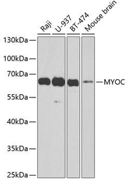
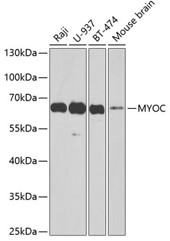
Anti-MYOC Antibody (CAB1589)
- SKU:
- CAB1589
- Product type:
- Antibody
- Reactivity:
- Human
- Reactivity:
- Mouse
- Host Species:
- Rabbit
- Isotype:
- IgG
- Antibody Type:
- Polyclonal Antibody
- Research Area:
- Cell Biology
Description
| 抗体名: | Anti-MYOC Antibody |
| 抗体コード: | CAB1589 |
| 抗体サイズ: | 20uL, 50uL, 100uL |
| 申し込み: | WB |
| 反応性: | Human, Mouse |
| 宿主種: | Rabbit |
| 免疫原: | Recombinant fusion protein containing a sequence corresponding to amino acids 245-504 of human MYOC (NP_000252.1). |
| 申し込み: | WB |
| 推奨希釈: | WB 1:500 - 1:2000 |
| 反応性: | Human, Mouse |
| ポジティブサンプル: | Raji, U-937, BT-474, Mouse brain |
| 免疫原: | Recombinant fusion protein containing a sequence corresponding to amino acids 245-504 of human MYOC (NP_000252.1). |
| 精製方法: | Affinity purification |
| ストレージバッファ: | Store at -20'C. Avoid freeze / thaw cycles. Buffer: PBS with 0.02% sodium azide, 50% glycerol, pH7.3. |
| アイソタイプ: | IgG |
| 順序: | CGEL VWVG EPLT LRTA ETIT GKYG VWMR DPKP TYPY TQET TWRI DTVG TDVR QVFE YDLI SQFM QGYP SKVH ILPR PLES TGAV VYSG SLYF QGAE SRTV IRYE LNTE TVKA EKEI PGAG YHGQ FPYS WGGY TDID LAVD EAGL WVIY STDE AKGA IVLS KLNP ENLE LEQT WETN IRKQ SVAN AFII CGTL YTVS SYTS ADAT VNFA YDTG TGIS KTLT IPFK NRYK YSSM IDYN PLEK KLFA WDNL NMVT YDIK LSKM |
| 遺伝子ID: | 4653 |
| Uniprot: | Q99972 |
| セルラーロケーション: | Cell projection, Cytoplasmic vesicle, Endoplasmic reticulum, Golgi apparatus, Mitochondrion, Mitochondrion inner membrane, Mitochondrion intermembrane space, Mitochondrion outer membrane, Rough endoplasmic reticulum, Secreted, cilium, exosome, extracellular matrix, extracellular space |
| 計算された分子量: | 56kDa |
| 観察された分子量: | 65kDa |
| 同義語: | MYOC, GLC1A, GPOA, JOAG, JOAG1, TIGR, myocilin |
| バックグラウンド: | MYOC encodes the protein myocilin, which is believed to have a role in cytoskeletal function. MYOC is expressed in many occular tissues, including the trabecular meshwork, and was revealed to be the trabecular meshwork glucocorticoid-inducible response protein (TIGR). The trabecular meshwork is a specialized eye tissue essential in regulating intraocular pressure, and mutations in MYOC have been identified as the cause of hereditary juvenile-onset open-angle glaucoma. |
| UniProt Protein Function: | MYOC: May participate in the obstruction of fluid outflow in the trabecular meshwork. Defects in MYOC are the cause of primary open angle glaucoma type 1A (GLC1A). Primary open angle glaucoma (POAG) is characterized by a specific pattern of optic nerve and visual field defects. The angle of the anterior chamber of the eye is open, and usually the intraocular pressure is increased. The disease is asymptomatic until the late stages, by which time significant and irreversible optic nerve damage has already taken place. Defects in MYOC are a cause of primary congenital glaucoma type 3A (GLC3A). An autosomal recessive form of primary congenital glaucoma (PCG). PCG is characterized by marked increase of intraocular pressure at birth or early choldhood, large ocular globes (buphthalmos) and corneal edema. It results from developmental defects of the trabecular meshwork and anterior chamber angle of the eye that prevent adequate drainage of aqueous humor. MYOC variations may contribute to GLC3A via digenic inheritance with CYP1B1 and/or another locus associated with the disease. |
| UniProt Protein Details: | Protein type:Secreted; Secreted, signal peptide; Endoplasmic reticulum Chromosomal Location of Human Ortholog: 1q23-q24 Cellular Component: extracellular matrix; Golgi apparatus; extracellular space; proteinaceous extracellular matrix; mitochondrial outer membrane; rough endoplasmic reticulum; endoplasmic reticulum; cytoplasmic membrane-bound vesicle; mitochondrial inner membrane; mitochondrial intermembrane space; cytoplasmic vesicle; cilium Molecular Function:protein binding; frizzled binding; fibronectin binding; myosin light chain binding; receptor tyrosine kinase binding Biological Process: clustering of voltage-gated sodium channels; negative regulation of stress fiber formation; negative regulation of cell-matrix adhesion; skeletal muscle hypertrophy; myelination in the peripheral nervous system; osteoblast differentiation; positive regulation of phosphoinositide 3-kinase cascade; positive regulation of protein kinase B signaling cascade; negative regulation of Rho protein signal transduction; positive regulation of focal adhesion formation; positive regulation of mitochondrial depolarization; regulation of MAPKKK cascade; positive regulation of stress fiber formation; neurite development; positive regulation of cell migration Disease: Glaucoma 1, Open Angle, A |
| NCBI Summary: | MYOC encodes the protein myocilin, which is believed to have a role in cytoskeletal function. MYOC is expressed in many occular tissues, including the trabecular meshwork, and was revealed to be the trabecular meshwork glucocorticoid-inducible response protein (TIGR). The trabecular meshwork is a specialized eye tissue essential in regulating intraocular pressure, and mutations in MYOC have been identified as the cause of hereditary juvenile-onset open-angle glaucoma. [provided by RefSeq, Jul 2008] |
| UniProt Code: | Q99972 |
| NCBI GenInfo Identifier: | 3024209 |
| NCBI Gene ID: | 4653 |
| NCBI Accession: | Q99972.2 |
| UniProt Secondary Accession: | Q99972,O00620, Q7Z6Q9, B2RD84, |
| UniProt Related Accession: | Q99972 |
| Molecular Weight: | 504 |
| NCBI Full Name: | Myocilin |
| NCBI Synonym Full Names: | myocilin, trabecular meshwork inducible glucocorticoid response |
| NCBI Official Symbol: | MYOC |
| NCBI Official Synonym Symbols: | GPOA; JOAG; TIGR; GLC1A; JOAG1; myocilin |
| NCBI Protein Information: | myocilin; myocilin 55 kDa subunit; mutated trabecular meshwork-induced glucocorticoid response protein |
| UniProt Protein Name: | Myocilin |
| UniProt Synonym Protein Names: | Myocilin 55 kDa subunit; Trabecular meshwork-induced glucocorticoid response proteinCleaved into the following 2 chains:Myocilin, N-terminal fragmentAlternative name(s):Myocilin 20 kDa N-terminal fragment |
| Protein Family: | Myocilin |
| UniProt Gene Name: | MYOC |
| UniProt Entry Name: | MYOC_HUMAN |


